- Home
- Objective
- Keynote Speakers
Mohamad Sawan Canada Research Chair in Smart Medical Devices Polystim Neurotechnologies Laboratory Polytechnique Montreal, Canada
Prof. Mohamad Sawan
Fabrice Wendling (PhD) holds a position of Director of Research at INSERM

Prof. fabrice Wendling
Metin Akay is currently the founding chair of the new Biomedical Engineering Department and the John S. Dunn professor of biomedical engineering at the University of Houston

Prof. Miten AKAY
Bart Bijnens

Prof. Bart Bijnens
ICREA Research Professor in the Department of Information and Communication Technologies at Universitat Pompeu Fabra (UPF)
Patrick Flandrin

Prof. Patrick Flandrin
CNRS "Research Director" at École normale supérieure de Lyon
Ghaleb Husseini Professor- AUS Chemical Eng. Department, Sharjah, UAE
 Prof. Ghaleb Husseini
Peter Stadler Algorithms, Computing in Mathematics, Natural Science, Engineering and Medicine, Bioinformatics University of Leipzig ,
 Prof. Peter Stadler
- Invited Speakers
- Partner Universities
- Lebanese University, LU, LB
- Saint Joseph University, USJ, LB
- Notre Dame University, NDU, LB
- Rafic Hariri University, RHU, LB
- Islamic Univ. of Lebanon,IUL, LB
- Saint Esprit of Kaslic Univ. USEK, LB
- More...
- Modality and submission
Paper presentation and submission

The conference will consist of oral presentations of fifteen minutes with few plenary sessions. All submitted papers will be peer reviewed. Accepted papers will be published as a collective work in an electronic format and will be submitted for inclusion to IEEE Xplore
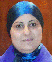
 Dorra Sellami Masmoudi
Professor, HdR, PhD, Ing,
Electrical Engineering Departmement
National Engineering School of Sfax, TunisiaUniversity of Sfax, ENIS, BP 1173 Sfax 3038 Tunisia
Title:Image Processing based Clinical Decision Support Systems for Breast Cancer Diagnosis
Abstract:
Breast cancer is becoming the last decade a serious health problem in different countries. It is the most frequent cancer affecting women. In 2012, the American cancer Society estimates that one in nine women in the USA can face breast cancer disease. In the European community, such a cancer covers 24 % of cancer cases. Mortality reduction by this disease is one of the main goals of scientists. It is becoming more and more significant owing to a wide spread of early investigations by screening tools for detection of abnormalities in their first stages of occurrence. Over the past two decades, many attempts have been made by scientists to assist the specialists in diagnosis of breast cancer, by developing computer-aided tools. Many screening modalities can be used, such as mammogram screening, echography, magnetic resonance imaging (MRI), positron emission tomography (PET), angio-mammogram screening, etc..Each image modality focuses on mapping on a 2D or 3D space a particular information related to the breast tissue, such as breast density, vascular information, dynamic characteristics of some cellules ( with use of a contrast agent), etc. Image processing of such modalities, in 2D as well as 3D representation, in that context, can have many vocations: First, it can be a processing aiming at highlighting abnormalities, sometimes slightly flooded, especially in dense breast. Accordingly, we should take the precaution of improving the contrast without distortion of the texture information, revealing in the diagnosis. Second, image processing can have a vocation of foreground segmentation of the Region Of Interest (abnormalities). Such segmentation should be of great accuracy, in order to preserve finest details of contours which are very pertinent for characterizing the benign/malign aspect of abnormalities. Microcalcification segmentation is a highly delicate step, since their tiny aspects. Once abnormality is delineated, another important step in image processing is characterization of shape and texture of the extracted abnormalities. We can also focus on volume reconstruction/characterization in a static representation as well as a dynamic representation from 3D MRI images. Feature extraction process can be appearance based, such as contour descriptors, since contour is a pertinent element interacting well in the nature (benign /malign) of the abnormality. Feature extraction can also exploit texture properties of malign abnormalities over benign ones. Global such as Zernike moments, LBP rotation invariant histograms of the image, Co-occurrence matrixes, as well as local features such as shape descriptors, can be adopted for feature extraction. Since most of the modalities carry complementary information such as tissue density (from mammography), vascularization of the abnormality (from MRI images), an important issue in medical processing is data fusion between a set of modalities of the same breast. Such fusion can be on a pixel level, for mapping information from different modalities on a same support, but fusion can be also done on feature level by choosing the most pertinent features for each modality. Some computer aided diagnosis systems complement the output processed by images, with some details related to the patient such as biopsy confirmation, hereditary factor, patient age, etc.. for making an overall classification of the studied case into the four Breast Imaging-Reporting and Data System (BI-RADS) categories. The BI-RADS classification system is rather an accurate precision of the type of the anomaly , accompanied by indicating the different future actions to be undertaken by the patient for a better prevention, such as a recommendation of biopsy or of a screening in coming few months, etc..
Key-words: Breast cancer, image processing, BIRADS, Computer aided diagnosis, mammogram, MRI, angiomammogram, PET, feature extraction, classificationBiography:
Dorra Sellami Masmoudi was born in Sfax, Tunisia in 1969. She received her engineer’s degree from Sfax National Engineering School in 1994 and has been awarded by the president of the republic of Tunisia. Subsequently, she joined IMS Laboratory at Bordeaux, in France and received her Ph.D. degree in 1998 and she has been assistant Professor at Sfax National Engineering School at 1999. Since 2006, she is a professor. From 2011, she is a full professor. Her researches include image and video processing, pattern recognition, Adaboost based detection, feature extraction, classification data fusion, neural and fuzzy based systems. It covers a large spectrum of applications such as medical image processing for automatic diagnosis of breast cancer, as well as biometry based personal identification via hand veins, fingerprint, iris, palmprint and face. Dorra Sellami Masmoudi supervised more than fiveteen PhD students. She is author of handreds of conference and journanl papers. She co-chaired two international conferences, the first in 2008 (IPTA’08) and the second in 2014 (IPAS’14).
- Keynote Speakers


 Mohamad Sawan